Menu

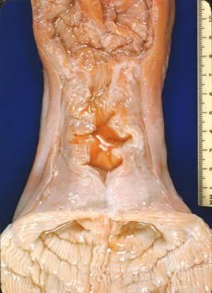
Abnormal cow reproductive organs
Credit: T. Y. Tanabe
Digital Credit: Harold Hafs
Publisher: None
Rights: No rights reserved - image is in the public domain
Description: This file contains five images of abnormal reproductive tracts from Dr. T. Y. Tanabe's research with cows at Pennsylvania State University in 1953 and 1954. Images 1 to 5 were digitized as they were captured originally. Labeling has been superimposed on the same images 1a to 5a (CC = cervical canal, CO = cervical os, Ov = ovary). The heifer in image 1 had two completely separate cervical canals, and no uterine body. In comparison, the cervix of the heifer illustrated in image 2 had two openings into the vaginal, but only one into the uterine body. Images 3 and 4 show heifers with one uterine horn disconnected from the body of the uterus. Heifers such as those in images 1, 2, 3, and 4 likely result from accidents of development late during the period of the embryo or early during the period of the fetus. Image 5, with unknown etiology, shows adhesions between the ovary and the infundibulum of the oviduct, completely blocking passage of ovulated oocytes.
Resolution: 1500x2076
File Size: 303.36 KB
