Menu

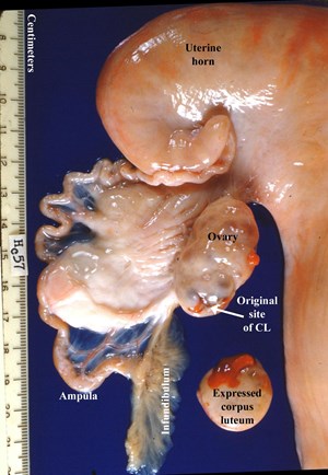
Manual expression of bovine corpus luteum
Credit: T. Y. Tanabe
Digital Credit: Harold Hafs
Publisher: None
Rights: No rights reserved - image is in the public domain
Description: This file contains seven images from Dr. T. Y. Tanabe's research on manual expression of bovine corpora lutea between 1950 and 1953 at Pennsylvania State University. Images 1 to 7 were digitized as they were captured originally. Labeling has been superimposed on the same images 1a to 7a (I = isthmus of oviduct, TUJ = tubouterine junction, CL = corpus luteum, CA = corpus albicans). The corpus luteum (CL) was a major target of research during the period of rapid expansion of artificial insemination of cattle. As surgical removal was a powerful method to study endocrine glands, researchers removed the CL through a surgical incision either in the flank or in the dorsal wall of the vagina. They could then follow changes in the cow's estrous cycle, and the excised CL could be studied in vitro. Alternatively, in the procedure used for the images in this file, Tanabe displaced the CL from the ovary by transrectal manual expression without invasive surgery. With no blood supply, the CL rapidly ceased to function and eventually was absorbed from the peritoneal cavity. Images 1, 2, 3 and 4 typify successful manual expression of CL; Tanabe recorded that the first three were done on day 12 after estrus. These images each illustrate the ovarian site where the CL had resided. The first three also show the CL that was recovered postmortem from the abdominal cavity 3 days later. Some hemorrhage was often found at the site where the CL had resided, as in image 2. At the same time he expressed CL on day 12, Tanabe typically observed a large follicle on one ovary, the cow began estrus about 2 to 4 days later, and a fertilized ovum could be recovered a few days following insemination during the induced estrus. Later, other researchers showed that follicles grow in two or three waves during the estrous cycle of cows. When the inhibitory influence of the CL is removed, the dominant follicle matures, and the cow begins estrus 2 to 4 days later and ovulates with normal fertility - as Tanabe observed. Images 5, 6, and 7 reveal that transrectal expression of CL can be problematic. Image 5 taken on day 5 after CL expression reveals a 2.5-cm diameter hematoma where the CL had resided. This cow was in estrus 2 days after CL expression, and had an ovulation point (OP) on the other ovary. While most cows show estrus 2 to 4 days after CL expression, the cow represented in image 6 began estrus 8 days later. In this cow, the superficial connection to the ovary was sufficient to maintain the CL. At 4 days after estrus, the same ovary had another CL emanating from the most recent ovulation. In image 7 at postmortem 12 days after CL expression, the periphery remained intact surrounding a blood clot where the central core had been located. Occasionally, transrectal expression of CL led to extensive intraperitoneal bleeding, and to adhesions between the infundibulum and the ovary. Most people abandoned surgical removal and trasrectal expression of CL after the discovery that prostaglandin F2a induced luteolysis.
Resolution: 1460x2110
File Size: 318.24 KB
