Menu

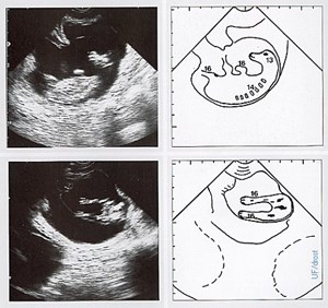
Bovine fetus, ultrasound at day 59
Credit: Pieterse MC (1999)
Digital Credit: Maarten Drost
Publisher: None
Rights: Name must appear as a credit whenever the image is used -
Description: Bovine fetus at 59 days of pregnancy. Transrectal ultrasonograms (left) of the fetus within the uterus, and explanatory diagrams (right) of the ultrasonograms; lateral view (top) and rear view (bottom). The amnionic vesicle becomes less turgid at this stage of pregnancy, permitting the fetus to be identified by palpation per rectum. The fetus was about the size of a mouse. Legend; 13 = head, 14 = trunk, 16 = limbs. This image is related to NAL #2296, 2298, 2299, and 2300.
Resolution: 478x450
File Size: 59.47 KB
