Menu

Brisket disease, cattle
Credit: R. J. Raleigh, Utah State University
Digital Credit: Melissa Foster
Publisher: American Institute of Nutrition
Rights: Image Gallery user terms
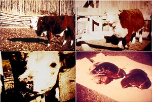
Description: These four images are of brisket disease in cattle, which is also known as high altitude disease or pulmonary hypertension, which is caused by elevated pulmonary arterial pressure. The elevated pressure is due to an oxygen shortage while at a high altitude. Low blood oxygen concentration causes hypoxic vasoconstriction (tightening of pulmonary arteries), leading to increased resistance to circulation. Since the blood is low in oxygen, any that enters the lungs is preferentially shunted to oxygenated areas. This causes resistance in blood flow in the arteries and the lungs. The heart then tries to compensate for this resistance by building up pressure to push the blood through. This causes enlargement of the right heart ventricle and the heart muscle may stretch so much that the valves inside do not meet, causing a backflow of blood with each contraction. Soon the built up pressure and back up of blood flow causes blood to pool in the lungs and fluid to leak out of the blood stream. The fluid is pushed into the air spaces (alveoli) and accumulates in the chest (brisket) causing brisket edema. The heart eventually cannot keep up leading to right heart dilation and/or heart failure. The first image in the upper left is of a two year old steer with a severe case of this disease. The earliest symptoms are listlessness, rough hair coat, dark profuse diarrhea, ascites (ascitic fluid leaks into the pleural cavity), and finally edema starting at the throat extending to the brisket and the body underline; in extreme cases to all lower extremities. Respiration is labored and pulse is weak, the jugular vein is frequently distended and showing pulsation. The mucous membranes are pale and nasal and ocular discharges may be seen. Animals may appear cyanotic and as the disease progresses, affected cattle become more reluctant to move and severely affected animals may collapse and die. The second image in the upper right is of a yearling heifer displaying similar effects as listed above. A calf is shown with fever blisters in the bottom left image. This symptom along with diarrhea is more common in calves than edema. In the last image, two livers are shown. The normal liver on the right and one from an animal that died of brisket disease on the left. Note the liver on the left is enlarged and discolored. The discoloration is due to chronic passive congestion and can cause severe lobular and vascular fibrosis. The lungs may have varying degrees of atelectasis (partial lung collapse), interstitial emphysema, edema, and pneumonia. In the heart, right ventricular hypertrophy, dilatation, and pulmonary arterial thrombosis can be seen.
Resolution: 5570x3730
File Size: 9.43 MB
